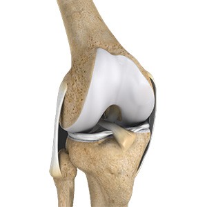
- Knee Anatomy
- Knee Conditions
- Knee Procedures
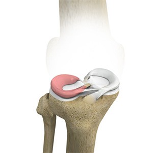
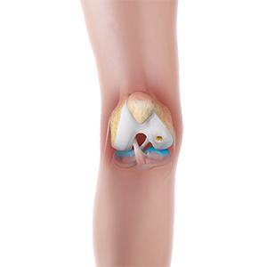
Lateral Meniscus Syndrome
The knee joint is formed by the union of two bones, namely the femur (thigh bone) and the tibia (lower leg bone). At the junction of these two bones is a cartilage called the meniscus, which acts as a shock absorber. There are two menisci – the lateral and medial menisci. The lateral meniscus is the outer meniscus of the knee joint and gives a cushioning effect during weight bearing activities.
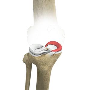
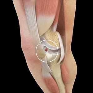
Medial Meniscus Syndrome
Medial meniscus syndrome refers to pain and dysfunction caused by injury or degeneration of the medial meniscus, the C-shaped cartilage on the inner side of the knee. Of the menisci within the knee, it is the medial that is more easily injured. Differences in the anatomical attachments of the medial meniscus compared to the lateral, mean that the medial meniscus becomes distorted during combined flexion and rotation movements in a manner not experienced on the lateral side.
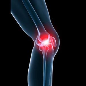
Knee Pain
Knee pain is a common condition affecting individuals of various age groups. It not only affects movement but also impacts your quality of life. An injury or disease of the knee joint or any structure surrounding the knee can result in knee pain. A precise diagnosis of the underlying cause is important to develop an appropriate treatment plan.
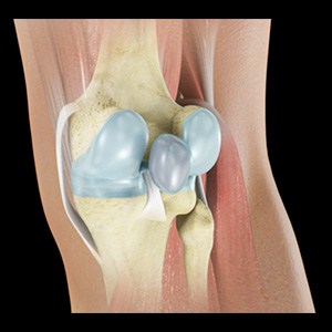
Baker’s Cyst
The knee consists of a fluid called synovial fluid, which reduces friction between the bones of the knee joint while you move your leg. Sometimes this fluid is produced in excess, resulting in its accumulation in the back of your knee. A Baker’s cyst or popliteal cyst is a fluid-filled swelling that develops into a lump behind the knee. This causes stiffness, tightness and pain behind your knee. It is commonly seen in women and people aged over 40 (although it can develop at any age).
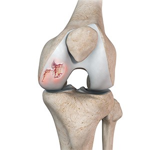
Osteochondritis Dissecans
Osteochondritis dissecans is a joint condition in which a piece of cartilage, along with a thin layer of the bone separates from the end of the bone because of inadequate blood supply. The separated fragments are sometimes called “joint mice”. These fragments may be localised, or may detach and fall into the joint space causing pain and joint instability.
ACL Tears
The anterior cruciate ligament (ACL) is one of the major ligaments of the knee. It is located in the middle of the knee and runs from the femur (thighbone) to the tibia (shinbone). The ACL prevents the tibia from sliding out in front of the femur. Together with the posterior cruciate ligament (PCL), it provides rotational stability to the knee.
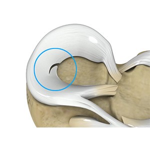
Meniscal Tears
A meniscal tear is a common knee injury in athletes, especially those involved in contact sports. A sudden bend or twist in your knee causes the meniscus to tear. Elderly people are more prone to degenerative meniscal tears as the cartilage wears out and weakens with age.
Ligament Injuries
The knee is a complex joint that consists of bone, cartilage, ligaments, and tendons that help in your joint’s movements. The knee is a hinge joint made up of two bones, the thighbone (femur) and shinbone (tibia). Ligaments are tough bands of tissue that connect one bone to another bone. The ligaments of the knee stabilize the knee joint. There are two important groups of ligaments that hold the bones of the knee joint together, collateral and cruciate ligaments - medial collateral ligament (MCL) and lateral collateral ligament (LCL), and anterior cruciate ligament (ACL) and posterior cruciate ligament (PCL).
Multiligament Instability
The knee is a complex joint of the body that is vital for movement. The four major ligaments of the knee are anterior cruciate ligament (ACL), posterior cruciate ligament (PCL), medial collateral ligament (MCL) and lateral collateral ligament (LCL). They play an important role in maintaining the stability of the knee. A multiligament injury is a tear in one or more ligaments of the knee, which affects the knee stability.
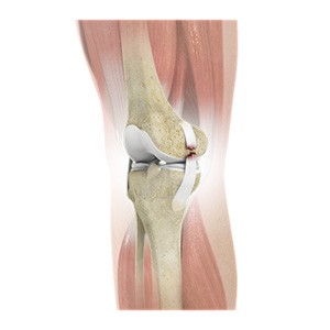
Knee Arthritis
The joint surface is covered by a smooth articular surface that allows pain-free movement in the joint. Arthritis is a general term covering numerous conditions where the joint surface or cartilage wears out. This surface can wear out for several reasons; often the definite cause is not known. Arthritis often affects the knee joint. When the articular cartilage wears out, the bone ends rub on one another and cause pain. The most common type of arthritis is osteoarthritis. It occurs with ageing and use.
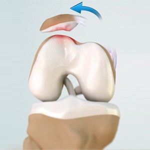
Patellar Dislocation / Patellofemoral Dislocation
Patellar dislocation occurs when the patella moves out of the patellofemoral groove, (trochlea) onto the bony head of the femur. If the kneecap partially comes out of the groove, it is called subluxation; if the kneecap completely comes out, it is called dislocation (luxation).
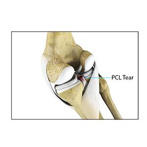
PCL Injuries
Posterior cruciate ligament (PCL), one of four major ligaments of the knee are situated at the back of the knee. It connects the thighbone (femur) to the shinbone (tibia). The PCL limits the backward motion of the shinbone. PCL injuries are very rare and are difficult to detect than other knee ligament injuries. Cartilage injuries, bone bruises, and ligament injuries often occur in combination with PCL injuries.
Chondral (Articular Cartilage Defects)
The articular or hyaline cartilage is the tissue lining the surface of the two bones in the knee joint. Cartilage helps the bones move smoothly against each other and can withstand the weight of your body during activities such as running and jumping. Articular cartilage does not have a direct blood supply to it, so has less capacity to repair itself. Once the cartilage is torn it will not heal easily and can lead to degeneration of the articular surface, leading to the development of osteoarthritis.
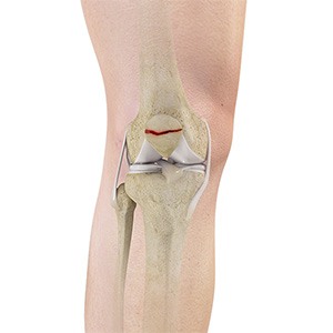
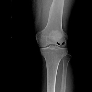
Patella Fracture
The kneecap or patella forms a part of the knee joint. It is present at the front of the knee, protecting the knee and providing attachment to various muscle groups of the thigh and leg. The undersurface of the kneecap and the lower end of the femur are coated with articular cartilage, which helps in smooth movement of the knee joint. A fracture in the kneecap is rare but common in adult males.
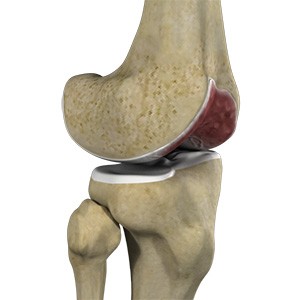
Osteonecrosis of the Knee
Osteonecrosis is a condition in which the death of a section of bone occurs because of lack of blood supply to it. It is one of the most common causes of knee pain in older women. Women over 60 years of age are commonly affected, three times more often than men.